| Henry Gray (1821–1865). Anatomy of the Human Body. 1918. |
| |
| 6d. 3. The Phalanges of the Foot |
| |
(Phalanges Digitorum Pedis)
The phalanges of the foot correspond, in number and general arrangement, with those of the hand; there are two in the great toe, and three in each of the other toes. They differ from them, however, in their size, the bodies being much reduced in length, and, especially in the first row, laterally compressed. | 1 |
| | | First Row.—The body of each is compressed from side to side, convex above, concave below. The base is concave; and the head presents a trochlear surface for articulation with the second phalanx. | 2 |
| | | Second Row.—The phalanges of the second row are remarkably small and short, but rather broader than those of the first row. | 3 |
| The ungual phalanges, in form, resemble those of the fingers; but they are smaller and are flattened from above downward; each presents a broad base for articulation with the corresponding bone of the second row, and an expanded distal extremity for the support of the nail and end of the toe. | 4 |
 |
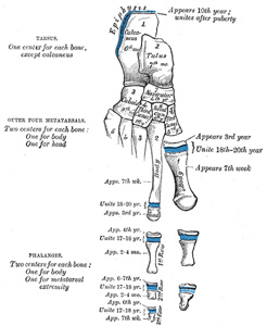
FIG. 289– Plan of ossification of the foot. (See enlarged image) | | |
| | | Articulations.—In the second, third, fourth, and fifth toes the phalanges of the first row articulate behind with the metatarsal bones, and in front with the second phalanges, which in their turn articulate with the first and third: the ungual phalanges articulate with the second. | 5 |
| | | Ossification of the Bones of the Foot (Fig. 289).—The tarsal bones are each ossified from a single center, excepting the calcaneus, which has an epiphysis for its posterior extremity. The centers make their appearance in the following order: calcaneus at the sixth month of fetal life; talus, about the seventh month; cuboid, at the ninth month; third cuneiform, during the first year; first cuneiform, in the third year; second cuneiform and navicular, in the fourth year. The epiphysis for the posterior extremity of the calcaneus appears at the tenth year, and unites with the rest of the bone soon after puberty. The posterior process of the talus is sometimes ossified from a separate center, and may remain distinct from the main mass of the bone, when it is named the os trigonum. | 6 |
| The metatarsal bones are each ossified from two centers: one for the body, and one for the head, of the second, third, fourth, and fifth metatarsals; one for the body, and one for the base, of the first metatarsal. 65 Ossification commences in the center of the body about the ninth week, and extends toward either extremity. The center for the base of the first metatarsal appears about the third year; the centers for the heads of the other bones between the fifth and eighth years; they join the bodies between the eighteenth and twentieth years. | 7 |
| The phalanges are each ossified from two centers: one for the body, and one for the base. The center for the body appears about the tenth week, that for the base between the fourth and tenth years; it joins the body about the eighteenth year. | 8 |
| Note 65. As was noted in the first metacarpal (see footnote, page 231), so in the first metatarsal, there is often a second epiphysis for its head. [back] |
|
|


