| Henry Gray (1821–1865). Anatomy of the Human Body. 1918. |
| |
| 5a. 3. The Frontal Bone |
| |
(Os Frontale)
The frontal bone resembles a cockle-shell in form, and consists of two portions—a vertical portion, the squama, corresponding with the region of the forehead; and an orbital or horizontal portion, which enters into the formation of the roofs of the orbital and nasal cavities. | 1 |
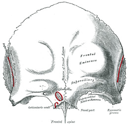
| | | Squama (squama frontalis).—Surfaces.—The external surface (Fig. 134) of this portion is convex and usually exhibits, in the lower part of the middle line, the remains of the frontal or metopic suture; in infancy this suture divides the bone into two, a condition which may persist throughout life. On either side of this suture, about 3 cm. above the supraorbital margin, is a rounded elevation, the frontal eminence (tuber frontale). These eminences vary in size in different individuals, are occasionally unsymmetrical, and are especially prominent in young skulls; the surface of the bone above them is smooth, and covered by the galea aponeurotica. Below the frontal eminences, and separated from them by a shallow groove, are two arched elevations, the superciliary arches; these are prominent medially, and are joined to one another by a smooth elevation named the glabella. They are larger in the male than in the female, and their degree of prominence depends to some extent on the size of the frontal air sinuses; 28 prominent ridges are, however, occasionally associated with small air sinuses. Beneath each superciliary arch is a curved and prominent margin, the supraorbital margin, which forms the upper boundary of the base of the orbit, and separates the squama from the orbital portion of the bone. The lateral part of this margin is sharp and prominent, affording to the eye, in that situation, considerable protection from injury; the medial part is rounded. At the junction of its medial and intermediate thirds is a notch, sometimes converted into a foramen, the supraorbital notch or foramen, which transmits the supraorbital vessels and nerve. A small aperture in the upper part of the notch transmits a vein from the diploë to join the supraorbital vein. The supraorbital margin ends laterally in the zygomatic process, which is strong and prominent, and articulates with the zygomatic bone. Running upward and backward from this process is a well-marked line, the temporal line, which divides into the upper and lower temporal lines, continuous, in the articulated skull, with the corresponding lines on the parietal bone. The area below and behind the temporal line forms the anterior part of the temporal fossa, and gives origin to the Temporalis muscle. Between the supraorbital margins the squama projects downward to a level below that of the zygomatic processes; this portion is known as the nasal part and presents a rough, uneven interval, the nasal notch, which articulates on either side of the middle line with the nasal bone, and laterally with the frontal process of the maxilla and with the lacrimal. The term nasion is applied to the middle of the frontonasal suture. From the center of the notch the nasal process projects downward and forward beneath the nasal bones and frontal processes of the maxillæ, and supports the bridge of the nose. The nasal process ends below in a sharp spine, and on either side of this is a small grooved surface which enters into the formation of the roof of the corresponding nasal cavity. The spine forms part of the septum of the nose, articulating in front with the crest of the nasal bones and behind with the perpendicular plate of the ethmoid. | 2 |
 |
FIG. 134– Frontal bone. Outer surface. (See enlarged image) | | |
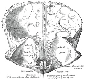
| The internal surface (Fig. 135) of the squama is concave and presents in the upper part of the middle line a vertical groove, the sagittal sulcus, the edges of which unite below to form a ridge, the frontal crest; the sulcus lodges the superior sagittal sinus, while its margins and the crest afford attachment to the falx cerebri. The crest ends below in a small notch which is converted into a foramen, the foramen cecum, by articulation with the ethmoid. This foramen varies in size in different subjects, and is frequently impervious; when open, it transmits a vein from the nose to the superior sagittal sinus. On either side of the middle line the bone presents depressions for the convolutions of the brain, and numerous small furrows for the anterior branches of the middle meningeal vessels. Several small, irregular fossæ may also be seen on either side of the sagittal sulcus, for the reception of the arachnoid granulations. | 3 |
| | | Orbital or Horizontal Part (pars orbitalis).—This portion consists of two thin triangular plates, the orbital plates, which form the vaults of the orbits, and are separated from one another by a median gap, the ethmoidal notch. | 4 |
 |
FIG. 135– Frontal bone. Inner surface. (See enlarged image) | | |
| | | Surfaces.—The inferior surface (Fig. 135) of each orbital plate is smooth and concave, and presents, laterally, under cover of the zygomatic process, a shallow depression, the lacrimal fossa, for the lacrimal gland; near the nasal part is a depression, the fovea trochlearis, or occasionally a small trochlear spine, for the attachment of the cartilaginous pulley of the Obliquus oculi superior. The superior surface is convex, and marked by depressions for the convolutions of the frontal lobes of the brain, and faint grooves for the meningeal branches of the ethmoidal vessels. | 5 |
| The ethmoidal notch separates the two orbital plates; it is quadrilateral, and filled, in the articulated skull, by the cribriform plate of the ethmoid. The margins of the notch present several half-cells which, when united with corresponding half-cells on the upper surface of the ethmoid, complete the ethmoidal air cells. Two grooves cross these edges transversely; they are converted into the anterior and posterior ethmoidal canals by the ethmoid, and open on the medial wall of the orbit. The anterior canal transmits the nasociliary nerve and anterior ethmoidal vessels, the posterior, the posterior ethmoidal nerve and vessels. In front of the ethmoidal notch, on either side of the frontal spine, are the openings of the frontal air sinuses. These are two irregular cavities, which extend backward, upward, and lateralward for a variable distance between the two tables of the skull; they are separated from one another by a thin bony septum, which often deviates to one or other side, with the result that the sinuses are rarely symmetrical. Absent at birth, they are usually fairly well-developed between the seventh and eighth years, but only reach their full size after puberty. They vary in size in different persons, and are larger in men than in women. 29 They are lined by mucous membrane, and each communicates with the corresponding nasal cavity by means of a passage called the frontonasal duct. | 6 |
| | | Borders.—The border of the squama is thick, strongly serrated, bevelled at the expense of the inner table above, where it rests upon the parietal bones, and at the expense of the outer table on either side, where it receives the lateral pressure of those bones; this border is continued below into a triangular, rough surface, which articulates with the great wing of the sphenoid. The posterior borders of the orbital plates are thin and serrated, and articulate with the small wings of the sphenoid. | 7 |
| | | Structure.—The squama and the zygomatic processes are very thick, consisting of diploic tissue contained between two compact laminæ; the diploic tissue is absent in the regions occupied by the frontal air sinuses. The orbital portion is thin, translucent, and composed entirely of compact bone; hence the facility with which instruments can penetrate the cranium through this part of the orbit; when the frontal sinuses are exceptionally large they may extend backward for a considerable distance into the orbital portion, which in such cases also consists of only two tables. | 8 |
| | | Ossification (Fig. 136).—The frontal bone is ossified in membrane from two primary centers, one for each half, which appear toward the end of the second month of fetal life, one above each supraorbital margin. From each of these centers ossification extends upward to form the corresponding half of the squama, and backward to form the orbital plate. The spine is ossified from a pair of secondary centers, on either side of the middle line; similar centers appear in the nasal part and zygomatic processes. At birth the bone consists of two pieces, separated by the frontal suture, which is usually obliterated, except at its lower part, by the eighth year, but occasionally persists throughout life. It is generally maintained that the development of the frontal sinuses begins at the end of the first or beginning of the second year, but Onodi’s researches indicate that development begins at birth. The sinuses are of considerable size by the seventh or eighth year, but do not attain their full proportions until after puberty. | 9 |
| | | Articulations.—The frontal articulates with twelve bones: the sphenoid, the ethmoid, the two parietals, the two nasals, the two maxillæ, the two lacrimals, and the two zygomatics. | 10 |
 |
FIG. 136– Frontal bone at birth. (See enlarged image) | | |
| Note 28. Some confusion is occasioned to students commencing the study of anatomy by the name “sinus” having been given to two different kinds of space connected with the skull. It may be as well, therefore, to state here that the “sinuses” in the interior of the cranium which produce the grooves on the inner surfaces of the bones are venous channels which convey the blood from the brain, while the “sinuses” external to the cranial cavity (the frontal, sphenoidal, ethmoidal, and maxillary) are hollow spaces in the bones themselves; they communicate with the nasal cavities and contain air. [back] |
| Note 29. Aldren Turner (The Accessory Sinuses of the Nose, 1901) gives the following measurements for a sinus of average size: height, 1 1/4 inches; breadth, 1 inch; depth from before backward, 1 inch. [back] |
|
|


