| Henry Gray (1821–1865). Anatomy of the Human Body. 1918. |
| |
| 2. The Digestive Apparatus |
| |
(Apparatus Digestorius; Organs Of Digestion)
The apparatus for the digestion of the food consists of the digestive tube and of certain accessory organs. | 1 |
| The Digestive Tube (alimentary canal) is a musculomembranous tube, about 9 metres long, extending from the mouth to the anus, and lined throughout its entire extent by mucous membrane. It has received different names in the various parts of its course: at its commencement is the mouth, where provision is made for the mechanical division of the food (mastication), and for its admixture with a fluid secreted by the salivary glands (insalivation); beyond this are the organs of deglutition, the pharynx and the esophagus, which convey the food into the stomach, in which it is stored for a time and in which also the first stages of the digestive process take place; the stomach is followed by the small intestine, which is divided for purposes of description into three parts, the duodenum, the jejunum, and ileum. In the small intestine the process of digestion is completed and the resulting products are absorbed into the blood and lacteal vessels. Finally the small intestine ends in the large intestine, which is made up of cecum, colon, rectum, and anal canal, the last terminating on the surface of the body at the anus. | 2 |
| The accessory organs are the teeth, for purposes of mastication; the three pairs of salivary glands—the parotid, submaxillary, and sublingual—the secretion from which mixes with the food in the mouth and converts it into a bolus and acts chemically on one of its constituents; the liver and pancreas, two large glands in the abdomen, the secretions of which, in addition to that of numerous minute glands in the walls of the alimentary canal, assist in the process of digestion. | 3 |
| | | The Development of the Digestive Tube.—The primitive digestive tube consists of two parts, viz.: (1) the fore-gut, within the cephalic flexure, and dorsal to the heart; and (2) the hind-gut, within the caudal flexure (Fig. 977). Between these is the wide opening of the yolk-sac, which is gradually narrowed and reduced to a small foramen leading into the vitelline duct. At first the fore-gut and hind-gut end blindly. The anterior end of the fore-gut is separated from the stomodeum by the buccopharyngeal membrane (Fig. 977); the hind-gut ends in the cloaca, which is closed by the cloacal membrane. | 4 |
 |
FIG. 977– Human embryo about fifteen days old. Brain and heart represented from right side. Digestive tube and yolk sac in median section. (After His.) (See enlarged image) | | |
| | | The Mouth.—The mouth is developed partly from the stomodeum, and partly from the floor of the anterior portion of the fore-gut. By the growth of the head end of the embryo, and the formation of the cephalic flexure, the pericardial area and the buccopharyngeal membrane come to lie on the ventral surface of the embryo. With the further expansion of the brain, and the forward bulging of the pericardium, the buccopharyngeal membrane is depressed between these two prominences. This depression constitutes the stomodeum (Fig. 977). It is lined by ectoderm, and is separated from the anterior end of the fore-gut by the buccopharyngeal membrane. This membrane is devoid of mesoderm, being formed by the apposition of the stomodeal ectoderm with the fore-gut entoderm; at the end of the third week it disappears, and thus a communication is established between the mouth and the future pharynx. No trace of the membrane is found in the adult; and the communication just mentioned must not be confused with the permanent isthmus faucium. The lips, teeth, and gums are formed from the walls of the stomodeum, but the tongue is developed in the floor of the pharynx. | 5 |
| The visceral arches extend in a ventral direction between the stomodeum and the pericardium; and with the completion of the mandibular arch and the formation of the maxillary processes, the mouth assumes the appearance of a pentagonal orifice. The orifice is bounded in front by the fronto-nasal process, behind by the mandibular arch, and laterally by the maxillary processes (Fig. 978). With the inward growth and fusion of the palatine processes (Figs. 50, 51), the stomodeum is divided into an upper nasal, and a lower buccal part. Along the free margins of the processes bounding the mouth cavity a shallow groove appears; this is termed the primary labial groove, and from the bottom of it a downgrowth of ectoderm takes place into the underlying mesoderm. The central cells of the ectodermal downgrowth degenerate and a secondary labial groove is formed; by the deepening of this, the lips and cheeks are separated from the alveolar processes of the maxillæ and mandible. | 6 |
| | | The Salivary Glands.—The salivary glands arise as buds from the epithelial lining of the mouth; the parotid appears during the fourth week in the angle between the maxillary process and the mandibular arch; the submaxillary appears in the sixth week, and the sublingual during the ninth week in the hollow between the tongue and the mandibular arch. | 7 |
 |
FIG. 978– Head end of human embryo of about thirty to thirty-one days. (From model by Peters.) (See enlarged image) | | |
 |
FIG. 979– Floor of pharynx of human embryo about twenty-six days old. (From model by Peters.) (See enlarged image) | | |
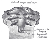 |
FIG. 980– Floor of pharynx of human embryo of about the end of the fourth week. (From model by Peters.) (See enlarged image) | | |
| | | The Tongue (Figs. 979 to 981).—The tongue is developed in the floor of the pharynx, and consists of an anterior or buccal and a posterior or pharyngeal part which are separated in the adult by the V-shaped sulcus terminalis. During the third week there appears, immediately behind the ventral ends of the two halves of the mandibular arch, a rounded swelling named the tuberculum impar, which was described by His as undergoing enlargement to form the buccal part of the tongue. More recent researches, however, show that this part of the tongue is mainly, if not entirely, developed from a pair of lateral swellings which rise from the inner surface of the mandibular arch and meet in the middle line. The tuberculum impar is said to form the central part of the tongue immediately in front of the foramen cecum, but Hammar insists that it is purely a transitory structure and forms no part of the adult tongue. From the ventral ends of the fourth arch there arises a second and larger elevation, in the center of which is a median groove or furrow. This elevation was named by His the furcula, and is at first separated from the tuberculum impar by a depression, but later by a ridge, the copula, formed by the forward growth and fusion of the ventral ends of the second and third arches. The posterior or pharyngeal part of the tongue is developed from the copula, which extends forward in the form of a V, so as to embrace between its two limbs the buccal part of the tongue. At the apex of the V a pit-like invagination occurs, to form the thyroid gland, and this depression is represented in the adult by the foramen cecum of the tongue. In the adult the union of the anterior and posterior parts of the tongue is marked by the V-shaped sulcus terminalis, the apex of which is at the foramen cecum, while the two limbs run lateralward and forward, parallel to, but a little behind, the vallate papillæ. | 8 |
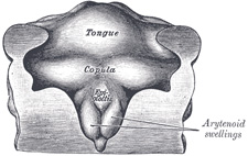 |
FIG. 981– Floor of pharynx of human embryo about thirty days old. (From model by Peter.) (See enlarged image) | | |
| | | The Palatine Tonsils.—The palatine tonsils are developed from the dorsal angles of the second branchial pouches. The entoderm which lines these pouches grows in the form of a number of solid buds into the surrounding mesoderm. These buds become hollowed out by the degeneration and casting off of their central cells, and by this means the tonsillar crypts are formed. Lymphoid cells accumulate around the crypts, and become grouped to form the lymphoid follicles; the latter, however, are not well-defined until after birth. | 9 |
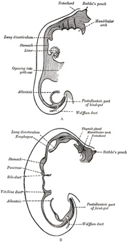 |
FIG. 982– Sketches in profile of two stages in the development of the human digestive tube. (His.) A X 30. B X 20. (See enlarged image) | | |
| | | The Further Development of the Digestive Tube.—The upper part of the fore-gut becomes dilated to form the pharynx (Fig. 977), in relation to which the branchial arches are developed (see page 65); the succeeding part remains tubular, and with the descent of the stomach is elongated to form the esophagus. About the fourth week a fusiform dilatation, the future stomach, makes its appearance, and beyond this the gut opens freely into the yolk-sac (Fig. 982, A and B). The opening is at first wide, but is gradually narrowed into a tubular stalk, the yolk-stalk or vitelline duct. Between the stomach and the mouth of the yolk-sac the liver diverticulum appears. From the stomach to the rectum the alimentary canal is attached to the notochord by a band of mesoderm, from which the common mesentery of the gut is subsequently developed. The stomach has an additional attachment, viz., to the ventral abdominal wall as far as the umbilicus by the septum transversum. The cephalic portion of the septum takes part in the formation of the diaphragm, while the caudal portion into which the liver grows forms the ventral mesogastrium (Fig. 984). The stomach undergoes a further dilatation, and its two curvatures can be recognized (Figs. 983, B, and 984), the greater directed toward the vertebral column and the lesser toward the anterior wall of the abdomen, while its two surfaces look to the right and left respectively. Behind the stomach the gut undergoes great elongation, and forms a V-shaped loop which projects downward and forward; from the bend or angle of the loop the vitelline duct passes to the umbilicus (Fig. 984). For a time a considerable part of the loop extends beyond the abdominal cavity into the umbilical cord, but by the end of the third month it is withdrawn within the cavity. With the lengthening of the tube, the mesoderm, which attaches it to the future vertebral column and carries the bloodvessels for the supply of the gut, is thinned and drawn out to form the posterior common mesentery. The portion of this mesentery attached to the greater curvature of the stomach is named the dorsal mesogastrium, and the part which suspends the colon is termed the mesocolon (Fig. 985). About the sixth week a diverticulum of the gut appears just behind the opening of the vitelline duct, and indicates the future cecum and vermiform process. The part of the loop on the distal side of the cecal diverticulum increases in diameter and forms the future ascending and transverse portions of the large intestine. Until the fifth month the cecal diverticulum has a uniform caliber, but from this time onward its distal part remains rudimentary and forms the vermiform process, while its proximal part expands to form the cecum. Changes also take place in the shape and position of the stomach. Its dorsal part or greater curvature, to which the dorsal mesogastrium is attached, grows much more rapidly than its ventral part or lesser curvature to which the ventral mesogastrium is fixed. Further, the greater curvature is carried downward and to the left, so that the right surface of the stomach is now directed backward and the left surface forward (Fig. 986), a change in position which explains why the left vagus nerve is found on the front, and the right vagus on the back of the stomach. The dorsal mesogastrium being attached to the greater curvature must necessarily follow its movements, and hence it becomes greatly elongated and drawn lateralward and ventralward from the vertebral column, and, as in the case of the stomach, the right surfaces of both the dorsal and ventral mesogastria are now directed backward, and the left forward. In this way a pouch, the bursa omentalis, is formed behind the stomach, and this increases in size as the digestive tube undergoes further development; the entrance to the pouch constitutes the future foramen epiploicum or foramen of Winslow. The duodenum is developed from that part of the tube which immediately succeeds the stomach; it undergoes little elongation, being more or less fixed in position by the liver and pancreas, which arise as diverticula from it. The duodenum is at first suspended by a mesentery, and projects forward in the form of a loop. The loop and its mesentery are subsequently displaced by the transverse colon, so that the right surface of the duodenal mesentery is directed backward, and, adhering to the parietal peritoneum, is lost. The remainder of the digestive tube becomes greatly elongated, and as a consequence the tube is coiled on itself, and this elongation demands a corresponding increase in the width of the intestinal attachment of the mesentery, which becomes folded. | 10 |
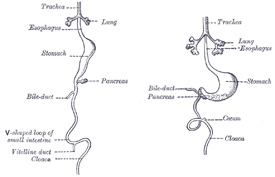 |
FIG. 983– Front view of two successive stages in the development of the digestive tube. (His.) (See enlarged image) | | |
 |
FIG. 984– The primitive mesentery of a six weeks’ human embryo, half schematic. (Kollmann.) (See enlarged image) | | |
 |
FIG. 985– Abdominal part of digestive tube and its attachment to the primitive or common mesentery. Human embryo of six weeks. (After Toldt.) (See enlarged image) | | |
| At this stage the small and large intestines are attached to the vertebral column by a common mesentery, the coils of the small intestine falling to the right of the middle line, while the large intestine lies on the left side. 158 | 11 |
| The gut is now rotated upon itself, so that the large intestine is carried over in front of the small intestine, and the cecum is placed immediately below the liver; about the sixth month the cecum descends into the right iliac fossa, and the large intestine forms an arch consisting of the ascending, transverse, and descending portions of the colon—the transverse portion crossing in front of the duodenum and lying just below the greater curvature of the stomach; within this arch the coils of the small intestine are disposed (Fig. 988). Sometimes the downward progress of the cecum is arrested, so that in the adult it may be found lying immediately below the liver instead of in the right iliac region. | 12 |
| Further changes take place in the bursa omentalis and in the common mesentery, and give rise to the peritoneal relations seen in the adult. The bursa omentalis, which at first reaches only as far as the greater curvature of the stomach, grows downward to form the greater omentum, and this downward extension lies in front of the transverse colon and the coils of the small intestine (Fig. 989). Above, before the pleuro-peritoneal opening is closed, the bursa omentalis sends up a diverticulum on either side of the esophagus; the left diverticulum soon disappears, but the right is constricted off and persists in most adults as a small sac lying within the thorax on the right side of the lower end of the esophagus. The anterior layer of the transverse mesocolon is at first distinct from the posterior layer of the greater omentum, but ultimately the two blend, and hence the greater omentum appears as if attached to the transverse colon (Fig. 990). The mesenteries of the ascending and descending parts of the colon disappear in the majority of cases, while that of the small intestine assumes the oblique attachment characteristic of its adult condition. | 13 |
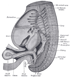 |
FIG. 986– Reconstruction of a human embryo of 17 mm. (After Mall.) (See enlarged image) | | |
| The lesser omentum is formed, as indicated above, by a thinning of the mesoderm or ventral mesogastrium, which attaches the stomach and duodenum to the anterior abdominal wall. By the subsequent growth of the liver this leaf of mesoderm is divided into two parts, viz., the lesser omentum between the stomach and liver, and the falciform and coronary ligaments between the liver and the abdominal wall and diaphragm (Fig. 989). | 14 |
 |
FIG. 987– Diagrams to illustrate two stages in the development of the digestive tube and its mesentery. The arrow indicates the entrance to the bursa omentalis. (See enlarged image) | | |
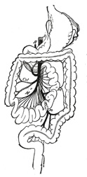 |
FIG. 988– Final disposition of the intestines and their vascular relations. (Jonnesco.) A. Aorta. H. Hepatic artery. M, Col. Branches of superior mesenteric artery. m, m’. Branches of inferior mesenteric artery. S. Splenic artery. (See enlarged image) | | |
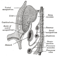 |
FIG. 989– Schematic figure of the bursa omentalis, etc. Human embryo of eight weeks. (Kollmann.) (See enlarged image) | | |
| | | The Rectum and Anal Canal.—The hind-gut is at first prolonged backward into the body-stalk as the tube of the allantois; but, with the growth and flexure of the tail-end of the embryo, the body-stalk, with its contained allantoic tube, is carried forward to the ventral aspect of the body, and consequently a bend is formed at the junction of the hind-gut and allantois. This bend becomes dilated into a pouch, which constitutes the entodermal cloaca; into its dorsal part the hind-gut opens, and from its ventral part the allantois passes forward. At a later stage the Wolffian and Müllerian ducts open into its ventral portion. The cloaca is, for a time, shut off from the anterior by a membrane, the cloacal membrane, formed by the apposition of the ectoderm and entoderm, and reaching, at first, as far forward as the future umbilicus. Behind the umbilicus, however, the mesoderm subsequently extends to form the lower part of the abdominal wall and symphysis pubis. By the growth of the surrounding tissues the cloacal membrane comes to lie at the bottom of a depression, which is lined by ectoderm and named the ectodermal cloaca (Fig. 991). | 15 |
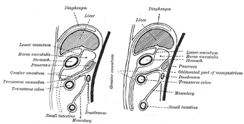 |
FIG. 990– Diagrams to illustrate the development of the greater omentum and transverse mesocolon. (See enlarged image) | | |
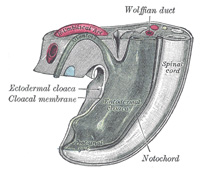 |
FIG. 991– Tail end of human embryo from fifteen to eighteen days old. (From model by Keibel.) (See enlarged image) | | |
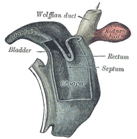 |
FIG. 992– Cloaca of human embryo from twenty-five to twenty-seven days old. (From model by Keibel.) (See enlarged image) | | |
| The entodermal cloaca is divided into a dorsal and a ventral part by means of a partition, the urorectal septum (Fig. 992), which grows downward from the ridge separating the allantoic from the cloacal opening of the intestine and ultimately fuses with the cloacal membrane and divides it into an anal and a urogenital part. The dorsal part of the cloaca forms the rectum, and the anterior part of the urogenital sinus and bladder. For a time a communication named the cloacal duct exists between the two parts of the cloaca below the urorectal septum; this duct occasionally persists as a passage between the rectum and urethra. The anal canal is formed by an invagination of the ectoderm behind the urorectal septum. This invagination is termed the proctodeum, and it meets with the entoderm of the hind-gut and forms with it the anal membrane. By the absorption of this membrane the anal canal becomes continuous with the rectum (Fig. 993). A small part of the hind-gut projects backward beyond the anal membrane; it is named the post-anal gut (Fig. 991), and usually becomes obliterated and disappears. 159 | 16 |
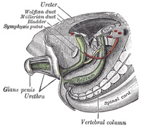 |
FIG. 993– Tail end of human embryo, from eight and a half to nine weeks old. (From model by Keibel.) (See enlarged image) | | |
| Note 158. Sometimes this condition persists throughout life, and it is then found that the duodenum does not cross from the right to the left side of the vertebral column, but lies entirely on the right side of the median plane, where it is continued into the jejunum; the arteries to the small intestine (aa. intestinales) also arise from the right instead of the left side of the superior mesenteric artery. [back] |
| Note 159. Consult, in this connection, the following article: “A Contribution to the Morphology of the Human Urinogenital Tract,” by D. Berry Hart, M.D., F.R.C.P.E., Journal of Anatomy and Physiology, April, 1901, vol. xxxv. [back] |
|
|


