| Henry Gray (1825–1861). Anatomy of the Human Body. 1918. |
FIG. 932
|

|
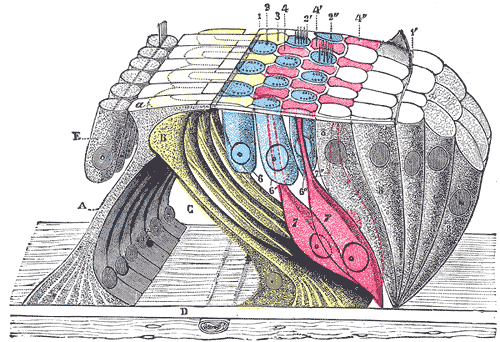
| The lamina reticularis and subjacent structures. (Schematic.) A. Internal rod of Corti, with a, its plate. B. External rod (in yellow). C. Tunnel of Corti. D. Membrana basilaris. E. Inner hair cells. 1, 1’. Internal and external borders of the membrana reticularis. 2, 2’, 2”. The three rows of circular holes (in blue). 3. First row of phalanges (in yellow). 4, 4’, 4”. Second, third, and fourth rows of phalanges (in red). 6, 6’, 6”. The three rows of outer hair cells (in blue). 7, 7’, 7”. Cells of Deiters. 8. Cells of Hensen and Claudius. (Testut.) |
|
|

