| Henry Gray (1821–1865). Anatomy of the Human Body. 1918. |
| |
| 5. Articulations of the Trunk. a. Articulations of the Vertebral Column |
| |
| These may be divided into the following groups, viz.: | 1 |
| I. Of the Vertebral Column. |
VI. Of the Cartilages of the Ribs with the
Sternum, and with Each Other. |
| II. Of the Atlas with the Axis. |
| III. Of the Vertebral Column with
the Cranium. |
VII. Of the Sternum. |
| IV. Of the Mandible. |
VIII. Of the Vertebral Column with the Pelvis. |
| V. Of the Ribs with the Vertebræ. |
IX. Of the Pelvis. |
|
| Articulations of the Vertebral Column
The articulations of the vertebral column consist of (1) a series of amphiarthrodial joints between the vertebral bodies, and (2) a series of diathrodial joints between the vertebral arches. | 2 |
| 1. Articulations of Vertebral Bodies (intercentral ligaments).—The articulations between the bodies of the vertebræ are amphiarthrodial joints, and the individual vertebræ move only slightly on each other. When, however, this slight degree of movement between the pairs of bones takes place in all the joints of the vertebral column, the total range of movement is very considerable. The ligaments of these articulations are the following: | 3 |
| The Anterior Longitudinal. |
The Posterior Longitudinal. |
| The Intervertebral Fibrocartilages. |
| | | | The Anterior Longitudinal Ligament (ligamentum longitudinale anterius; anterior common ligament) (Figs. 301, 312).—The anterior longitudinal ligament is a broad and strong band of fibers, which extends along the anterior surfaces of the bodies of the vertebræ, from the axis to the sacrum. It is broader below than above,
thicker in the thoracic than in the cervical and lumbar regions, and somewhat thicker opposite the bodies of the vertebræ than opposite the intervertebral fibrocartilages. It is attached, above, to the body of the axis, where it is continuous with the anterior atlantoaxial ligament, and extends down as far as the upper part of the front of the sacrum. It consists of dense longitudinal fibers, which are intimately adherent to the intervertebral fibrocartilages and the prominent margins of the vertebræ, but not to the middle parts of the bodies. In the latter situation the ligament is thick and serves to fill up the concavities on the anterior surfaces, and to make the front of the vertebral column more even. It is composed of several layers of fibers, which vary in length, but are closely interlaced with each other. The most superficial fibers are the longest and extend between four or five vertebræ. A second, subjacent set extends between two or three vertebræ while a third set, the shortest and deepest, reaches from one vertebra to the next. At the sides of the bodies the ligament consists of a few short fibers which pass from one vertebra to the next, separated from the concavities of the vertebral bodies by oval apertures for the passage of vessels. | 4 |
 |
FIG. 301– Median sagittal section of two lumbar vertebræ and their ligaments. (See enlarged image) | | |
| | | The Posterior Longitudinal Ligament (ligamentum longitudinale posterius; posterior common ligament) (Figs. 301, 302).—The posterior longitudinal ligament is situated within the vertebral canal, and extends along the posterior surfaces of the bodies of the vertebræ, from the body of the axis, where it is continuous with the membrana tectoria, to the sacrum. It is broader above than below, and thicker in the thoracic than in the cervical and lumbar regions. In the situation of the intervertebral fibrocartilages and contiguous margins of the vertebræ, where the ligament is more intimately adherent, it is broad, and in the thoracic and lumbar regions presents a series of dentations with intervening concave margins; but it is narrow and thick over the centers of the bodies, from which it is separated by the basivertebral veins. This ligament is composed of smooth, shining, longitudinal fibers, denser and more compact than those of the anterior ligament, and consists of superficial layers occupying the interval between three or four vertebræ, and deeper layers which extend between adjacent vertebræ. | 5 |
| | | The Intervertebral Fibrocartilages (fibrocartilagines intervertebrales; intervertebral disks) (Figs. 301, 313).—The intervertebral fibrocartilages are interposed between the adjacent surfaces of the bodies of the vertebræ, from the axis to the sacrum, and form the chief bonds of connection between the vertebræ. They vary in shape, size, and thickness, in different parts of the vertebral column. In shape and size they correspond with the surfaces of the bodies between which they are placed, except in the cervical region, where they are slightly smaller from side to side than the corresponding bodies. In thickness they vary not only in the different regions of the column, but in different parts of the same fibrocartilage; they are thicker in front than behind in the cervical and lumbar regions, and thus contribute to the anterior convexities of these parts of the column; while they are of nearly uniform thickness in the thoracic region, the anterior concavity of this part of the column being almost entirely owing to the shape of the vertebral bodies. The intervertebral fibrocartilages constitute about one-fourth of the length of the vertebral column, exclusive of the first two vertebræ; but this amount is not equally distributed between the various bones, the cervical and lumbar portions having, in proportion to their length, a much greater amount than the thoracic region, with the result that these parts possess greater pliancy and freedom of movement. The intervertebral fibrocartilages are adherent, by their surfaces, to thin layers of hyaline cartilage which cover the upper and under surfaces of the bodies of the vertebræ; in the lower cervical vertebræ, however, small joints lined by synovial membrane are occasionally present between the upper surfaces of the bodies and the margins of the fibrocartilages on either side. By their circumferences the intervertebral fibrocartilages are closely connected in front to the anterior, and behind to the posterior, longitudinal ligaments. In the thoracic region they are joined laterally, by means of the interarticular ligaments, to the heads of those ribs which articulate with two vertebræ. | 6 |
 |
FIG. 302– Posterior longitudinal ligament, in the thoracic region. (See enlarged image) | | |
| | | Structure of the Intervertebral Fibrocartilages.—Each is composed, at its circumference, of laminæ of fibrous tissue and fibrocartilage, forming the annulus fibrosus; and, at its center, of a soft, pulpy, highly elastic substance, of a yellowish color, which projects considerably above the surrounding level when the disk is divided horizontally. This pulpy substance (nucleus pulposus), especially well-developed in the lumbar region, is the remains of the notochord. The laminæ are arranged concentrically; the outermost consist of ordinary fibrous tissue, the others of white fibrocartilage. The laminæ are not quite vertical in their direction, those near the circumference being curved outward and closely approximated; while those nearest the center curve in the opposite direction, and are somewhat more widely separated. The fibers of which each lamina is composed are directed, for the most part, obliquely from above downward, the fibers of adjacent laminæ passing in opposite directions and varying in every layer; so that the fibers of one layer are directed across those of another, like the limbs of the letter X. This laminar arrangement belongs to about the outer half of each fibrocartilage. The pulpy substance presents no such arrangement, and consists of a fine fibrous matrix, containing angular cells united to form a reticular structure. | 7 |
| The intervertebral fibrocartilages are important shock absorbers. Under pressure the highly elastic nucleus pulposus becomes flatter and broader and pushes the more resistant fibrous laminæ outward in all directions. | 8 |
| 2. Articulations of Vertebral Arches.—The joints between the articular processes of the vertebræ belong to the arthrodial variety and are enveloped by capsules lined by synovial membranes; while the laminæ, spinous and transverse processes are connected by the following ligaments: | 9 |
| The Ligamenta Flava. |
|
The Ligamentum Nuchæ. |
| The Supraspinal. |
|
The Interspinal. |
| The Intertransverse. | | | | | The Articular Capsules (capsulæ articulares; capsular ligaments) (Fig. 301).—The articular capsules are thin and loose, and are attached to the margins of the articular processes of adjacent vertebræ. They are longer and looser in the cervical than in the thoracic and lumbar regions. | 10 |
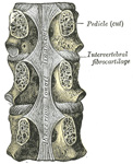
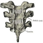
| | | The Ligamenta Flava (ligamenta subflava, Fig. 303).—The ligamenta flava connect the laminæ of adjacent vertebræ, from the axis to the first segment of the sacrum. They are best seen from the interior of the vertebral canal; when looked at from the outer surface they appear short, being overlapped by the laminæ. Each ligament consists of two lateral portions which commence one on either side of the roots of the articular processes, and extend backward to the point where the laminæ meet to form the spinous process; the posterior margins of the two portions are in contact and to a certain extent united, slight intervals being left for the passage of small vessels. Each consists of yellow elastic tissue, the fibers of which, almost perpendicular in direction, are attached to the anterior surface of the lamina above, some distance from its inferior margin, and to the posterior surface and upper margin of the lamina below. In the cervical region the ligaments are thin, but broad and long; they are thicker in the thoracic region, and thickest in the lumbar region. Their marked elasticity serves to preserve the upright posture, and to assist the vertebral column in resuming it after flexion. | 11 |
 |
FIG. 303– Vertebral arches of three thoracic vertebræ viewed from the front. (See enlarged image) | | |
| | | The Supraspinal Ligament (ligamentum supraspinale; supraspinous ligament) (Fig. 301).—The supraspinal ligament is a strong fibrous cord, which connects together the apices of the spinous processes from the seventh cervical vertebra to the sacrum; at the points of attachment to the tips of the spinous processes fibrocartilage is developed in the ligament. It is thicker and broader in the lumbar than in the thoracic region, and intimately blended, in both situations, with the neighboring fascia. The most superficial fibers of this ligament extend over three or four vertebræ; those more deeply seated pass between two or three vertebræ while the deepest connect the spinous processes of neighboring vertebræ. Between the spinous processes it is continuous with the interspinal ligaments. It is continued upward to the external occipital protuberance and median nuchal line, as the ligamentum nuchæ. | 12 |
| | | The Ligamentum Nuchæ.—The ligamentum nuchæ is a fibrous membrane, which, in the neck, represents the supraspinal ligaments of the lower vertebræ. It extends from the external occipital protuberance and median nuchal line to the spinous process of the seventh cervical vertebra. From its anterior border a fibrous lamina is given off, which is attached to the posterior tubercle of the atlas, and to the spinous processes of the cervical vertebræ, and forms a septum between the muscles
on either side of the neck. In man it is merely the rudiment of an important elastic ligament, which, in some of the lower animals, serves to sustain the weight of the head. | 13 |
| | | The Interspinal Ligaments (ligamenta interspinalia; interspinous ligaments) (Fig. 301).—The interspinal ligaments thin and membranous, connect adjoining spinous processes and extend from the root to the apex of each process. They meet the ligamenta flava in front and the supraspinal ligament behind. They are narrow and elongated in the thoracic region; broader, thicker, and quadrilateral in form in the lumbar region; and only slightly developed in the neck. | 14 |
| | | The Intertransverse Ligaments (ligamenta intertransversaria).—The intertransverse ligaments are interposed between the transverse processes. In the cervical region they consist of a few irregular, scattered fibers; in the thoracic region they are rounded cords intimately connected with the deep muscles of the back; in the lumbar region they are thin and membranous. | 15 |
| | | Movements.—The movements permitted in the vertebral column are: flexion, extension, lateral movement, circumduction, and rotation. | 16 |
| In flexion, or movement forward, the anterior longitudinal ligament is relaxed, and the intervertebral fibrocartilages are compressed in front; while the posterior longitudinal ligament, the ligamenta flava, and the inter- and supraspinal ligaments are stretched, as well as the posterior fibers of the intervertebral fibrocartilages. The interspaces between the laminæ are widened, and the inferior articular processes glide upward, upon the superior articular processes of the subjacent vertebræ. Flexion is the most extensive of all the movements of the vertebral column, and is freest in the lumbar region. | 17 |
| In extension, or movement backward, an exactly opposite disposition of the parts takes place. This movement is limited by the anterior longitudinal ligament, and by the approximation of the spinous processes. It is freest in the cervical region. | 18 |
| In lateral movement, the sides of the intervertebral fibrocartilages are compressed, the extent of motion being limited by the resistance offered by the surrounding ligaments. This movement may take place in any part of the column, but is freest in the cervical and lumbar regions. | 19 |
| Circumduction is very limited, and is merely a succession of the preceding movements. | 20 |
| Rotation is produced by the twisting of the intervertebral fibrocartilages; this, although only slight between any two vertebræ, allows of a considerable extent of movement when it takes place in the whole length of the column, the front of the upper part of the column being turned to one or other side. This movement occurs to a slight extent in the cervical region, is freer in the upper part of the thoracic region, and absent in the lumbar region. | 21 |
| The extent and variety of the movements are influenced by the shape and direction of the articular surfaces. In the cervical region the upward inclination of the superior articular surfaces allows of free flexion and extension. Extension can be carried farther than flexion; at the upper end of the region it is checked by the locking of the posterior edges of the superior atlantal facets in the condyloid fossæ of the occipital bone; at the lower end it is limited by a mechanism whereby the inferior articular processes of the seventh cervical vertebra slip into grooves behind and below the superior articular processes of the first thoracic. Flexion is arrested just beyond the point where the cervical convexity is straightened; the movement is checked by the apposition of the projecting lower lips of the bodies of the vertebræ with the shelving surfaces on the bodies of the subjacent vertebræ. Lateral flexion and rotation are free in the cervical region; they are, however, always combined. The upward and medial inclinations of the superior articular surfaces impart a rotary movement during lateral flexion, while pure rotation is prevented by the slight medial slope of these surfaces. | 22 |
| In the thoracic region, notably in its upper part, all the movements are limited in order to reduce interference with respiration to a minimum. The almost complete absence of an upward inclination of the superior articular surfaces prohibits any marked flexion, while extension is checked by the contact of the inferior articular margins with the laminæ, and the contact of the spinous processes with one another. The mechanism between the seventh cervical and the first thoracic vertebræ, which limits extension of the cervical region, will also serve to limit flexion of the thoracic region when the neck is extended. Rotation is free in the thoracic region: the superior articular processes are segments of a cylinder whose axis is in the mid-ventral line of the vertebral bodies. The direction of the articular facets would allow of free lateral flexion, but this movement is considerably limited in the upper part of the region by the resistance of the ribs and sternum. | 23 |
| In the lumbar region flexion and extension are free. Flexion can be carried farther than extension, and is possible to just beyond the straightening of the lumbar curve; it is, therefore, greatest at the lowest part where the curve is sharpest. The inferior articular facets are not in close apposition with the superior facets of the subjacent vertebræ, and on this account a considerable amount of lateral flexion is permitted. For the same reason a slight amount of rotation can be carried out, but this is so soon checked by the interlocking of the articular surfaces that it is negligible. | 24 |
| The principal muscles which produce flexion are the Sternocleidomastoideus, Longus capitis, and Longus colli; the Scaleni; the abdominal muscles and the Psoas major. Extension is produced by the intrinsic muscles of the back, assisted in the neck by the Splenius, Semispinales dorsi and cervicis, and the Multifidus. Lateral motion is produced by the intrinsic muscles of the back by the Splenius, the Scaleni, the Quadratus lumborum, and the Psoas major, the muscles of one side only acting; and rotation by the action of the following muscles of one side only, viz., the Sternocleidomastoideus, the Longus capitis, the Scaleni, the Multifidus, the Semispinalis capitis, and the abdominal muscles. | 25 |
|
|


