| Henry Gray (1821–1865). Anatomy of the Human Body. 1918. |
III. Syndesmology |
| |
| |
| Introduction |
| |
| THE BONES of the skeleton are joined to one another at different parts of their surfaces, and such connections are termed Joints or Articulations. Where the joints are immovable, as in the articulations between practically all the bones of the skull, the adjacent margins of the bones are almost in contact, being separated merely by a thin layer of fibrous membrane, named the sutural ligament. In certain regions at the base of the skull this fibrous membrane is replaced by a layer of cartilage. Where slight movement combined with great strength is required, the osseous surfaces are united by tough and elastic fibrocartilages, as in the joints between the vertebral bodies, and in the interpubic articulation. In the freely movable joints the surfaces are completely separated; the bones forming the articulation are expanded for greater convenience of mutual connection, covered by cartilage and enveloped by capsules of fibrous tissue. The cells lining the interior of the fibrous capsule form an imperfect membrane—the synovial membrane—which secretes a lubricating fluid. The joints are strengthened by strong fibrous bands called ligaments, which extend between the bones forming the joint. | 1 |
| | | Bone.—Bone constitutes the fundamental element of all the joints. In the long bones, the extremities are the parts which form the articulations; they are generally somewhat enlarged; and consist of spongy cancellous tissue with a thin coating of compact substance. In the flat bones, the articulations usually take place at the edges; and in the short bones at various parts of their surfaces. The layer of compact bone which forms the joint surface, and to which the articular cartilage is attached, is called the articular lamella. It differs from ordinary bone tissue in that it contains no Haversian canals, and its lacunæ are larger and have no canaliculi. The vessels of the cancellous tissue, as they approach the articular lamella, turn back in loops, and do not perforate it; this layer is consequently denser and firmer than ordinary bone, and is evidently designed to form an unyielding support for the articular cartilage. | 2 |
| | | Cartilage.—Cartilage is a non-vascular structure which is found in various parts of the body—in adult life chiefly in the joints, in the parietes of the thorax, and in various tubes, such as the trachea and bronchi, nose, and ears, which require to be kept permanently open. In the fetus, at an early period, the greater part of the skeleton is cartilaginous; as this cartilage is afterward replaced by bone, it is called temporary, in contradistinction to that which remains unossified during the whole of life, and is called permanent. | 3 |
| Cartilage is divided, according to its minute structure, into hyaline cartilage, white fibrocartilage, and yellow or elastic fibrocartilage. | 4 |
| | | Hyaline Cartilage.—Hyaline cartilage consists of a gristly mass of a firm consistence, but of considerable elasticity and pearly bluish color. Except where it coats the articular ends of bones, it is covered externally by a fibrous membrane, the perichondrium, from the vessels of which it imbibes its nutritive fluids, being itself destitute of bloodvessels. It contains no nerves. Its intimate structure is very simple. If a thin slice be examined under the microscope, it will be found to consist of cells of a rounded or bluntly angular form, lying in groups of two or more in a granular or almost homogeneous matrix (Fig. 292). The cells, when arranged in groups of two or more, have generally straight outlines where they are in contact with each other, and in the rest of their circumference are rounded. They consist of clear translucent protoplasm in which fine interlacing filaments and minute granules are sometimes present; imbedded in this are one or two round nuclei, having the usual intranuclear network. The cells are contained in cavities in the matrix, called cartilage lacunæ; around these the matrix is arranged in concentric lines, as if it had been formed in sucessive portions around the cartilage cells. This constitutes the so-called capsule of the space. Each lacuna is generally occupied by a single cell, but during the division of the cells it may contain two, four, or eight cells. | 5 |
 |
FIG. 292– Human cartilage cells from the cricoid cartilage. (See enlarged image) | | |
| The matrix is transparent and apparently without structure, or else presents a dimly granular appearance, like ground glass. Some observers have shown that the matrix of hyaline cartilage, and especially of the articular variety, after prolonged maceration, can be broken up into fine fibrils. These fibrils are probably of the same nature, chemically, as the white fibers of connective tissue. It is believed by some histologists that the matrix is permeated by a number of fine channels, which connect the lacunæ with each other, and that these canals communicate with the lymphatics of the perichondrium, and thus the structure is permeated by a current of nutrient fluid. | 6 |
| Articular cartilage, costal cartilage, and temporary cartilage are all of the hyaline variety. They present differences in the size, shape, and arrangement of their cells. | 7 |
 |
FIG. 293– Vertical section of articular cartilage. (See enlarged image) | | |
 |
FIG. 294– Costal cartilage from a man, aged seventy-six years, showing the development of fibrous structure in the matrix. In several portions of the specimen two or three generations of cells are seen enclosed in a parent cell wall. Highly magnified. (See enlarged image) | | |
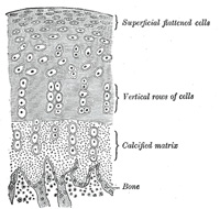
| In Articular Cartilage (Fig. 293), which shows no tendency to ossification, the matrix is finely granular; the cells and nuclei are small, and are disposed parallel to the surface in the superficial part, while nearer to the bone they are arranged in vertical rows. Articular cartilages have a tendency to split in a vertical direction; in disease this tendency becomes very manifest. The free surface of articular cartilage, where it is exposed to friction, is not covered by perichondrium, although a layer of connective tissue continuous with that of the synovial membrane can be traced in the adult over a small part of its circumference, and here the cartilage cells are more or less branched and pass insensibly into the branched connective tissue corpuscles of the synovial membrane. Articular cartilage forms a thin incrustation upon the joint surfaces of the bones, and its elasticity enables it to break the force of concussions, while its smoothness affords ease and freedom of movement. It varies in thickness according to the shape of the articular surface on which it lies; where this is convex the cartilage is thickest at the center, the reverse being the case on concave articular surfaces. It appears to derive its nutriment partly from the vessels of the neighboring synovial membrane and partly from those of the bone upon which it is implanted. Toynbee has shown that the minute vessels of the cancellous tissue as they approach the articular lamella dilate and form arches, and then return into the substance of the bone. | 8 |
| In Costal Cartilage the cells and nuclei are large, and the matrix has a tendency to fibrous striation, especially in old age (Fig. 294). In the thickest parts of the costal cartilages a few large vascular channels may be detected. This appears, at first sight, to be an exception to the statement that cartilage is a non-vascular tissue, but is not so really, for the vessels give no branches to the cartilage substance itself, and the channels may rather be looked upon as involutions of the perichondrium. The xiphoid process and the cartilages of the nose, larynx, and trachea (except the epiglottis and corniculate cartilages of the larynx, which are composed of elastic fibrocartilage) resemble the costal cartilages in microscopic characteristics. The arytenoid cartilage of the larynx shows a transition from hyaline cartilage at its base to elastic cartilage at the apex. | 9 |
| The hyaline cartilages, especially in adult and advanced life, are prone to calcify—that is to say, to have their matrix permeated by calcium salts without any appearance of true bone. The process of calcification occurs frequently, in such cartilages as those of the trachea and in the costal cartilages, where it may be succeeded by conversion into true bone. | 10 |
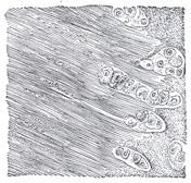
| | | White Fibrocartilage.—White fibrocartilage consists of a mixture of white fibrous tissue and cartilaginous tissue in various proportions; to the former of these constituents it owes its flexibility and toughness, and to the latter its elasticity. When examined under the microscope it is found to be made up of fibrous connective tissue arranged in bundles, with cartilage cells between the bundles; the cells to a certain extent resemble tendon cells, but may be distinguished from them by being surrounded by a concentrically striated area of cartilage matrix and by being less flattened (Fig. 295). The white fibrocartilages admit of arrangement into four groups—interarticular, connecting, circumferential, and stratiform. | 11 |
 |
FIG. 295– White fibrocartilage from an intervertebral fibrocartilage. (See enlarged image) | | |
| 1. The Interarticular Fibrocartilages (menisci) are flattened fibrocartilaginous plates, of a round, oval, triangular, or sickle-like form, interposed between the articular cartilages of certain joints. They are free on both surfaces, usually thinner toward the center than at the circumference, and held in position by the attachment of their margins and extremities to the surrounding ligaments. The synovial membranes of the joints are prolonged over them. They are found in the temporomandibular, sternoclavicular, acromioclavicular, wrist, and knee joints—i. e., in those joints which are most exposed to violent concussion and subject to frequent movement. Their uses are to obliterate the intervals between opposed surfaces in their various motions; to increase the depths of the articular surfaces and give ease to the gliding movements; to moderate the effects of great pressure and deaden the intensity of the shocks to which the parts may be subjected. Humphry has pointed out that these interarticular fibrocartilages serve an important purpose in increasing the varieties of movement in a joint. Thus in the knee joint there are two kinds of motion, viz., angular movement and rotation, although it is a hinge joint, in which, as a rule, only one variety of motion is permitted; the former movement takes place between the condyles of the femur and the interarticular cartilages, the latter between the cartilages and the head of the tibia. So, also, in the temporomandibular joint, the movements of opening and shutting the mouth take place between the fibrocartilage and the mandible, the grinding movement between the mandibular fossa and the fibrocartilage, the latter moving with the mandible. | 12 |
| 2. The Connecting Fibrocartilages are interposed between the bony surfaces of those joints which admit of only slight mobility, as between the bodies of the vertebræ. They form disks which are closely adherent to the opposed surfaces. Each disk is composed of concentric rings of fibrous tissue, with cartilaginous laminæ interposed, the former tissue predominating toward the circumference, the latter toward the center. | 13 |
| 3. The Circumferential Fibrocartilages consist of rims of fibrocartilage, which surround the margins of some of the articular cavities, e. g., the glenoidal labrum of the hip, and of the shoulder; they serve to deepen the articular cavities and to protect their edges. | 14 |
| 4. The Stratiform Fibrocartilages are those which form a thin coating to osseous grooves through which the tendons of certain muscles glide. Small masses of fibrocartilage are also developed in the tendons of some muscles, where they glide over bones, as in the tendons of the Peronæus longus and Tibialis posterior. | 15 |
| The distinguishing feature of cartilage chemically is that it yields on boiling a substance called chondrin, very similar to gelatin, but differing from it in several of its reactions. It is now believed that chondrin is not a simple body, but a mixture of gelatin with mucinoid substances, chief among which, perhaps, is a compound termed chondro-mucoid. | 16 |
| | | Ligaments.—Ligaments are composed mainly of bundles of white fibrous tissue placed parallel with, or closely interlaced with one another, and present a white, shining, silvery appearance. They are pliant and flexible, so as to allow perfect freedom of movement, but strong, tough, and inextensible, so as not to yield readily to applied force. Some ligaments consist entirely of yellow elastic tissue, as the ligamenta flava which connect together the laminæ of adjacent vertebræ, and the ligamentum nuchæ in the lower animals. In these cases the elasticity of the ligament is intended to act as a substitute for muscular power. | 17 |
| | | The Articular Capsules.—The articular capsules form complete envelopes for the freely movable joints. Each capsule consists of two strata—an external (stratum fibrosum) composed of white fibrous tissue, and an internal (stratum synoviale) which is a secreting layer, and is usually described separately as the synovial membrane. | 18 |
| The fibrous capsule is attached to the whole circumference of the articular end of each bone entering into the joint, and thus entirely surrounds the articulation. | 19 |
| The synovial membrane invests the inner surface of the fibrous capsule, and is reflected over any tendons passing through the joint cavity, as the tendon of the Popliteus in the knee, and the tendon of the Biceps brachii in the shoulder. It is composed of a thin, delicate, connective tissue, with branched connective-tissue corpuscles. Its secretion is thick, viscid, and glairy, like the white of an egg, and is hence termed synovia. In the fetus this membrane is said, by Toynbee, to be continued over the surfaces of the cartilages; but in the adult such a continuation is wanting, excepting at the circumference of the cartilage, upon which it encroaches for a short distance and to which it is firmly attached. In some of the joints the synovial membrane is thrown into folds which pass across the cavity; they are especially distinct in the knee. In other joints there are flattened folds, subdivided at their margins into fringe-like processes which contain convoluted vessels. These folds generally project from the synovial membrane near the margin of the cartilage, and lie flat upon its surface. They consist of connective tissue, covered with endothelium, and contain fat cells in variable quantities, and, more rarely, isolated cartilage cells; the larger folds often contain considerable quantities of fat. | 20 |
| Closely associated with synovial membrane, and therefore conveniently described in this section, are the mucous sheaths of tendons and the mucous bursæ. | 21 |
| Mucous sheaths (vaginæ mucosæ) serve to facilitate the gliding of tendons in fibroösseous canals. Each sheath is arranged in the form of an elongated closed sac, one layer of which adheres to the wall of the canal, and the other is reflected upon the surface of the enclosed tendon. These sheaths are chiefly found surrounding the tendons of the Flexor and Extensor muscles of the fingers and toes as they pass through fibroösseous canals in or near the hand and foot. | 22 |
| Bursæ mucosæ are interposed between surfaces which glide upon each other. They consist of closed sacs containing a minute quantity of clear viscid fluid, and may be grouped, according to their situations, under the headings subcutaneous, submuscular, subfacial, and subtendinous | 23 |
|
|


