| Henry Gray (1821–1865). Anatomy of the Human Body. 1918. |
| |
| 4b. 4. The Ulnar Artery |
| |
(A. Ulnaris)
The ulnar artery (Fig. 528), the larger of the two terminal branches of the brachial, begins a little below the bend of the elbow, and, passing obliquely downward, reaches the ulnar side of the forearm at a point about midway between the elbow and the wrist. It then runs along the ulnar border to the wrist, crosses the transverse carpal ligament on the radial side of the pisiform bone, and immediately beyond this bone divides into two branches, which enter into the formation of the superficial and deep volar arches. | 1 |
| | | Relations.—(a) In the forearm.—In its upper half, it is deeply seated, being covered by the Pronator teres, Flexor carpi radialis, Palmaris longus, and Flexor digitorum sublimis; it lies upon the Brachialis and Flexor digitorum profundus. The median nerve is in relation with the medial side of the artery for about 2.5 cm. and then crosses the vessel, being separated from it by the ulnar head of the Pronator teres. In the lower half of the forearm it lies upon the Flexor digitorum profundus, being covered by the integument and the superficial and deep fasciæ, and placed between the Flexor carpi ulnaris and Flexor digitorum sublimis. It is accompanied by two venæ comitantes, and is overlapped in its middle third by the Flexor carpi ulnaris; the ulnar nerve lies on the medial side of the lower two-thirds of the artery, and the palmar cutaneous branch of the nerve descends on the lower part of the vessel to the palm of the hand. | 2 |
| (b) At the wrist (Fig. 527) the ulnar artery is covered by the integument and the volar carpal ligament, and lies upon the transverse carpal ligament. On its medial side is the pisiform bone, and, somewhat behind the artery, the ulnar nerve. | 3 |
| | | Peculiarities.—The ulnar artery varies in its origin in the proportion of about one in thirteen cases; it may arise about 5 to 7 cm. below the elbow, but more frequently higher, the brachial being more often the source of origin than the axillary. Variations in the position of this vessel are more common than in the radial. When its origin is normal, the course of the vessel is rarely changed. When it arises high up, it is almost invariably superficial to the Flexor muscles in the forearm, lying commonly beneath the fascia, more rarely between the fascia and integument. In a few cases, its position was subcutaneous in the upper part of the forearm, and subaponeurotic in the lower part. | 4 |
| | | Branches.—The branches of the ulnar artery may be arranged in the following groups: | 5 |
| In the Forearm |
Anterior Recurrent. |
At the Wrist |
Volar Carpal. |
| Posterior Recurrent. |
Dorsal Carpal. |
| Common Interosseous. |
In the Hand |
Deep Volar. |
| Muscular. |
Superficial Volar Arch. |
|
| The anterior ulnar recurrent artery (a. recurrentes ulnaris anterior) arises immediately below the elbow-joint, runs upward between the Brachialis and Pronator teres, supplies twigs to those muscles, and, in front of the medial epicondyle, anastomoses with the superior and inferior ulnar collateral arteries. | 6 |
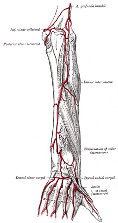
| The posterior ulnar recurrent artery (a. recurrentes ulnaris posterior) is much larger, and arises somewhat lower than the preceding. It passes backward and medialward on the Flexor digitorum profundus, behind the Flexor digitorum sublimis, and ascends behind the medial epicondyle of the humerus. In the interval between this process and the olecranon, it lies beneath the Flexor carpi ulnaris, and ascending between the heads of that muscle, in relation with the ulnar nerve, it supplies the neighboring muscles and the elbow-joint, and anastomoses with the superior and inferior ulnar collateral and the interosseous recurrent arteries (Fig. 529). | 7 |
| The common interosseous artery (a. interossea communis) (Fig. 528), about 1 cm. in length, arises immediately below the tuberosity of the radius, and, passing backward to the upper border of the interosseous membrane, divides into two branches, the volar and dorsal interosseous arteries. | 8 |
| The Volar Interosseous Artery (a. interossea volaris; anterior interosseous artery) (Fig. 528), passes down the forearm on the volar surface of the interosseous membrane. It is accompanied by the volar interosseous branch of the median nerve, and overlapped by the contiguous margins of the Flexor digitorum profundus and Flexor pollicis longus, giving off in this situation muscular branches, and the nutrient arteries of the radius and ulna. At the upper border of the Pronator quadratus it pierces the interosseous membrane and reaches the back of the forearm, where it anastomoses with the dorsal interosseous artery (Fig. 529). It then descends, in company with the terminal portion of the dorsal interosseous nerve, to the back of the wrist to join the dorsal carpal net-work. The volar interosseous artery gives off a slender branch, the arteria mediana, which accompanies the median nerve, and gives offsets to its substance; this artery is sometimes much enlarged, and runs with the nerve into the palm of the hand. Before it pierces the interosseous membrane the volar interosseous sends a branch downward behind the Pronator quadratus to join the volar carpal network. | 9 |
 |
FIG. 529– Arteries of the back of the forearm and hand. (See enlarged image) | | |
| The Dorsal Interosseous Artery (a. interossea dorsalis; posterior interosseous artery) (Fig. 529) passes backward between the oblique cord and the upper border of the interosseous membrane. It appears between the contiguous borders of the Supinator and the Abductor pollicis longus, and runs down the back of the forearm between the superficial and deep layers of muscles, to both of which it distributes branches. Where it lies upon the Abductor pollicis longus and the Extensor pollicis brevis, it is accompanied by the dorsal interosseous nerve. At the lower part of the forearm it anastomoses with the termination of the volar interosseous artery, and with the dorsal carpal network. It gives off, near its origin, the interosseous recurrent artery, which ascends to the interval between the lateral epicondyle and olecranon, on or through the fibers of the Supinator, but beneath the Anconæus, and anastomoses with the radial collateral branch of the profunda brachii, the posterior ulnar recurrent and the inferior ulnar collateral. | 10 |
| The muscular branches (rami musculares) are distributed to the muscles along the ulnar side of the forearm. | 11 |
| The volar carpal branch (ramus carpeus volares; anterior ulnar carpal artery) is a small vessel which crosses the front of the carpus beneath the tendons of the Flexor digitorum profundus, and anastomoses with the corresponding branch of the radial artery. | 12 |
| The dorsal carpal branch (ramus carpeus dorsalis; posterior ulnar carpal artery) arises immediately above the pisiform bone, and winds backward beneath the tendon of the Flexor carpi ulnaris; it passes across the dorsal surface of the carpus beneath the Extensor tendons, to anastomose with a corresponding branch of the radial artery. Immediately after its origin, it gives off a small branch, which runs along the ulnar side of the fifth metacarpal bone, and supplies the ulnar side of the dorsal surface of the little finger. | 13 |
| The deep volar branch (ramus volaris profundus; profunda branch) (Fig. 528) passes between the Abductor digiti quinti and Flexor digiti quinti brevis and through the origin of the Opponens digiti quinti; it anastomoses with the radial artery, and completes the deep volar arch. | 14 |
| The superficial volar arch (arcus volaris superficialis; superficial palmar arch) (Fig. 527) is formed by the ulnar artery, and is usually completed by a branch from the a. volaris indicis radialis, but sometimes by the superficial volar or by a branch from the a. princeps pollicis of the radial artery. The arch passes across the palm, describing a curve, with its convexity downward. | 15 |
| | | Relations.—The superficial volar arch is covered by the skin, the Palmaris brevis, and the palmar aponeurosis. It lies upon the transverse carpal ligament, the Flexor digiti quinti brevis and Opponens digiti quinti, the tendons of the Flexor digitorum sublimis, the Lumbricales, and the divisions of the median and ulnar nerves. | 16 |
| Three Common Volar Digital Arteries (aa. digitales volares communes; palmar digital arteries) (Fig. 527) arise from the convexity of the arch and proceed downward on the second, third, and fourth Lumbricales. Each receives the corresponding volar metacarpal artery and then divides into a pair of proper volar digital arteries (aa. digitales volares propriæ; collateral digital arteries) which run along the contiguous sides of the index, middle, ring, and little fingers, behind the corresponding digital nerves; they anastomose freely in the subcutaneous tissue of the finger tips and by smaller branches near the interphalangeal joints. Each gives off a couple of dorsal branches which anastomose with the dorsal digital arteries, and supply the soft parts on the back of the second and third phalanges, including the matrix of the finger-nail. The proper volar digital artery for medial side of the little finger springs from the ulnar artery under cover of the Palmaris brevis. | 17 |
|
|


