| Henry Gray (1821–1865). Anatomy of the Human Body. 1918. |
| |
| 3a. 4. The Internal Carotid Artery |
| |
(A. Carotis Interna)
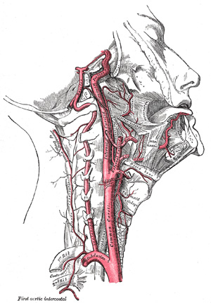
The internal carotid artery (Fig. 513) supplies the anterior part of the brain, the eye and its appendages, and sends branches to the forehead and nose. Its size, in the adult, is equal to that of the external carotid, though, in the child, it is larger than that vessel. It is remarkable for the number of curvatures that it presents in different parts of its course. It occasionally has one or two flexures near the base of the skull, while in its passage through the carotid canal and along the side of the body of the sphenoid bone it describes a double curvature and resembles the italic letter S. | 1 |
 |
FIG. 513– The internal carotid and vertebral arteries. Right side. (See enlarged image) | | |
| | | Course and Relations.—In considering the course and relations of this vessel it may be divided into four portions: cervical, petrous, cavernous, and cerebral. | 2 |
| | | Cervical Portion.—This portion of the internal carotid begins at the bifurcation of the common carotid, opposite the upper border of the thyroid cartilage, and runs perpendicularly upward, in front of the transverse processes of the upper three cervical vertebræ, to the carotid canal in the petrous portion of the temporal bone. It is comparatively superficial at its commencement, where it is contained in the carotid triangle, and lies behind and lateral to the external carotid, overlapped by the Sternocleidomastoideus, and covered by the deep fascia, Platysma, and integument: it then passes beneath the parotid gland, being crossed by the hypoglossal nerve, the Digastricus and Stylohyoideus, and the occipital and posterior auricular arteries. Higher up, it is separated from the external carotid by the Styloglossus and Stylopharyngeus, the tip of the styloid process and the stylohyoid ligament, the glossopharyngeal nerve and the pharyngeal branch of the vagus. It is in relation, behind, with the Longus capitis, the superior cervical ganglion of the sympathetic trunk, and the superior laryngeal nerve; laterally, with the internal jugular vein and vagus nerve, the nerve lying on a plane posterior to the artery; medially, with the pharynx, superior laryngeal nerve, and ascending pharyngeal artery. At the base of the skull the glossopharyngeal, vagus, accessory, and hypoglossal nerves lie between the artery and the internal jugular vein. | 3 |
| | | Petrous Portion.—When the internal carotid artery enters the canal in the petrous portion of the temporal bone, it first ascends a short distance, then curves forward and medialward, and again ascends as it leaves the canal to enter the cavity of the skull between the lingula and petrosal process of the sphenoid. The artery lies at first in front of the cochlea and tympanic cavity; from the latter cavity it is separated by a thin, bony lamella, which is cribriform in the young subject, and often partly absorbed in old age. Farther forward it is separated from the semilunar ganglion by a thin plate of bone, which forms the floor of the fossa for the ganglion and the roof of the horizontal portion of the canal. Frequently this bony plate is more or less deficient, and then the ganglion is separated from the artery by fibrous membrane. The artery is separated from the bony wall of the carotid canal by a prolongation of dura mater, and is surrounded by a number of small veins and by filaments of the carotid plexus, derived from the ascending branch of the superior cervical ganglion of the sympathetic trunk. | 4 |
| | | Cavernous Portion.—In this part of its course, the artery is situated between the layers of the dura mater forming the cavernous sinus, but covered by the lining membrane of the sinus. It at first ascends toward the posterior clinoid process, then passes forward by the side of the body of the sphenoid bone, and again curves upward on the medial side of the anterior clinoid process, and perforates the dura mater forming the roof of the sinus. This portion of the artery is surrounded by filaments of the sympathetic nerve, and on its lateral side is the abducent nerve. | 5 |
| | | Cerebral Portion.—Having perforated the dura mater on the medial side of the anterior clinoid process, the internal carotid passes between the optic and oculomotor nerves to the anterior perforated substance at the medial extremity of the lateral cerebral fissure, where it gives off its terminal or cerebral branches. | 6 |
| | | Peculiarities.—The length of the internal carotid varies according to the length of the neck, and also according to the point of bifurcation of the common carotid. It arises sometimes from the arch of the aorta; in such rare instances, this vessel has been found to be placed nearer the middle line of the neck than the external carotid, as far upward as the larynx, when the latter vessel crossed the internal carotid. The course of the artery, instead of being straight, may be very tortuous. A few instances are recorded in which this vessel was altogether absent; in one of these the common carotid passed up the neck, and gave off the usual branches of the external carotid; the cranial portion of the internal carotid was replaced by two branches of the internal maxillary, which entered the skull through the foramen rotundum and foramen ovale, and joined to form a single vessel. | 7 |
| | | Branches.—The cervical portion of the internal carotid gives off no branches. Those from the other portions are: | 8 |
| From the Petrous Portion |
Caroticotympanic. |
| Artery of the Pterygoid Canal. |
|
From the Cavernous Portion |
Cavernous. |
| Hypophyseal. |
| Semilunar. |
| Anterior Meningeal. |
| Ophthalmic. |
|
| From the Cerebral Portion |
Anterior Cerebral. |
| Middle Cerebral. |
| Posterior Communicating. |
| Choroidal. | |
| 1. The caroticotympanic branch (ramus caroticotympanicus; tympanic branch) is small; it enters the tympanic cavity through a minute foramen in the carotid canal, and anastomoses with the anterior tympanic branch of the internal maxillary, and with the stylomastoid artery. | 9 |
| 2. The artery of the pterygoid canal (a. canilis pterygoidei [Vidii]; Vidian artery) is a small, inconstant branch which passes into the pterygoid canal and anastomoses with a branch of the internal maxillary artery. | 10 |
| 3. The cavernous branches are numerous small vessels which supply the hypophysis, the semilunar ganglion, and the walls of the cavernous and inferior petrosal sinuses. Some of them anastomose with branches of the middle meningeal. | 11 |
| 4. The hypophyseal branches are one or two minute vessels supplying the hypophysis. | 12 |
| 5. The semilunar branches are small vessels to the semilunar ganglion. | 13 |
| 6. The anterior meningeal branch (a. meningea anterior) is a small branch which passes over the small wing of the sphenoid to supply the dura mater of the anterior cranial fossa; it anastomoses with the meningeal branch from the posterior ethmoidal artery. | 14 |
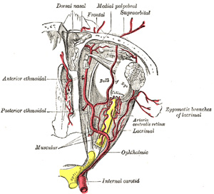
| 7. The ophthalmic artery (a. ophthalmica) (Fig. 514) arises from the internal carotid, just as that vessel is emerging from the cavernous sinus, on the medial side of the anterior clinoid process, and enters the orbital cavity through the optic foramen, below and lateral to the optic nerve. It then passes over the nerve to reach the medial wall of the orbit, and thence horizontally forward, beneath the lower border of the Obliquus superior, and divides it into two terminal branches, the frontal and dorsal nasal. As the artery crosses the optic nerve it is accompanied by the nasociliary nerve, and is separated from the frontal nerve by the Rectus superior and Levator palpebræ superioris. | 15 |
| | | Branches.—The branches of the ophthalmic artery may be divided into an orbital group, distributed to the orbit and surrounding parts; and an ocular group, to the muscles and bulb of the eye. | 16 |
| Orbital Group. |
| Ocular Group. |
| Lacrimal. |
| Central Artery of the Retina. |
| Supraorbital. |
| Short Posterior Ciliary. |
| Posterior Ethmoidal. |
| Long Posterior Ciliary. |
| Anterior Ethmoidal. |
| Anterior Ciliary. |
| Medial Palpebral. |
| Muscular. |
| Frontal. |
|
|
| Dorsal Nasal. |
|
| |
| The Lacrimal Artery (a. lacrimalis) arises close to the optic foramen, and is one of the largest branches derived from the ophthalmic: not infrequently it is given off before the artery enters the orbit. It accompanies the lacrimal nerve along the upper border of the Rectus lateralis, and supplies the lacrimal gland. Its terminal branches, escaping from the gland, are distributed to the eyelids and conjunctiva: of those supplying the eyelids, two are of considerable size and are named the lateral palpebral arteries; they run medialward in the upper and lower lids respectively and anastomose with the medial palpebral arteries, forming an arterial circle in this situation. The lacrimal artery give off one or two zygomatic branches, one of which passes through the zygomatico-temporal foramen, to reach the temporal fossa, and anastomoses with the deep temporal arteries; another appears on the cheek through the zygomatico-facial foramen, and anastomoses with the transverse facial. A recurrent branch passes backward through the lateral part of the superior orbital fissure to the dura mater, and anastomoses with a branch of the middle meningeal artery. The lacrimal artery is sometimes derived from one of the anterior branches of the middle meningeal artery. | 17 |
 |
FIG. 514– The ophthalmic artery and its branches. (See enlarged image) | | |
| The Supraorbital Artery (a. supraorbitalis) springs from the ophthalmic as that vessel is crossing over the optic nerve. It passes upward on the medial borders of the Rectus superior and Levator palpebræ, and meeting the supraorbital nerve accompanies it between the periosteum and Levator palpebræ to the supraorbital foramen; passing through this it divides into a superficial and a deep branch, which supply the integument, the muscles, and the pericranium of the forehead, anastomosing with the frontal, the frontal branch of the superficial temporal, and the artery of the opposite side. This artery in the orbit supplies the Rectus superior and the Levator palpebræ, and sends a branch across the pulley of the Obliquus superior, to supply the parts at the medial palpebral commissure. At the supraorbital foramen it frequently transmits a branch to the diploë. | 18 |
| The Ethmoidal Arteries are two in number: posterior and anterior. The posterior ethmoidal artery, the smaller, passes through the posterior ethmoidal canal, supplies the posterior ethmoidal cells, and, entering the cranium, gives off a meningeal branch to the dura mater, and nasal branches which descend into the nasal cavity through apertures in the cribriform plate, anastomosing with branches of the sphenopalatine. The anterior ethmoidal artery accompanies the nasociliary nerve through the anterior ethmoidal canal, supplies the anterior and middle ethmoidal cells and frontal sinus, and, entering the cranium, gives off a meningeal branch to the dura mater, and nasal branches; these latter descend into the nasal cavity through the slit by the side of the crista galli, and, running along the groove on the inner surface of the nasal bone, supply branches to the lateral wall and septum of the nose, and a terminal branch which appears on the dorsum of the nose between the nasal bone and the lateral cartilage. | 19 |
 |
FIG. 515– Bloodvessels of the eyelids, front view. 1, supraorbital artery and vein; 2, nasal artery; 3, angular artery, the terminal branch of 4, the facial artery; 5, suborbital artery; 6, anterior branch of the superficial temporal artery; 6’, malar branch of the transverse artery of the face; 7, lacrimal artery; 8, superior palpebral artery with 8’, its external arch; 9, anastomoses of the superior palpebral with the superficial temporal and lacrimal; 10, inferior palpebral artery; 11, facial vein; 12, angular vein; 13, branch of the superficial temporal vein. (Testut.) (See enlarged image) | | |
| The Medial Palpebral Arteries (aa. palpebrales mediales; internal palpebral arteries), two in number, superior and inferior, arise from the ophthalmic, opposite the pulley of the Obliquus superior; they leave the orbit to encircle the eyelids near their free margins, forming a superior and an inferior arch, which lie between the Orbicularis oculi and the tarsi. The superior palpebral anastomoses, at the lateral angle of the orbit, with the zygomaticoörbital branch of the temporal artery and with the upper of the two lateral palpebral branches from the lacrimal artery; the inferior palpebral anastomoses, at the lateral angle of the orbit, with the lower of the two lateral palpebral branches from the lacrimal and with the transverse facial artery, and, at the medial part of the lid, with a branch from the angular artery. From this last anastomoses a branch passes to the nasolacrimal duct, ramifying in its mucous membrane, as far as the inferior meatus of the nasal cavity. | 20 |
| The Frontal Artery (a. frontalis), one of the terminal branches of the ophthalmic, leaves the orbit at its medial angle with the supratrochlear nerve, and, ascending on the forehead, supplies the integument, muscles, and pericranium, anastomosing with the supraorbital artery, and with the artery of the opposite side. | 21 |
| The Dorsal Nasal Artery (a. dorsalis nasi; nasal artery), the other terminal branch of the ophthalmic, emerges from the orbit above the medial palpebral ligament, and, after giving a twig to the upper part of the lacrimal sac, divides into two branches, one of which crosses the root of the nose, and anastomoses with the angular artery, the other runs along the dorsum of the nose, supplies its outer surface; and anastomoses with the artery of the opposite side, and with the lateral nasal branch of the external maxillary. | 22 |
| The Central Artery of the Retina (a. centralis retinœ) is the first and one of the smallest branches of the ophthalmic artery. It runs for a short distance within the dural sheath of the optic nerve, but about 1.25 cm. behind the eyeball it pierces the nerve obliquely, and runs forward in the center of its substance to the retina. Its mode of distribution will be described with the anatomy of the eye. | 23 |
| The Ciliary Arteries (aa. ciliares) are divisible into three groups, the long and short, posterior, and the anterior. The short posterior ciliary arteries from six to twelve in number, arise from the ophthalmic, or its branches; they pass forward around the optic nerve to the posterior part of the eyeball, pierce the sclera around the entrance of the nerve, and supply the choroid and ciliary processes. The long posterior ciliary arteries, two in number, pierce the posterior part of the sclera at some little distance from the optic nerve, and run forward, along either side of the eyeball, between the sclera and choroid, to the ciliary muscle, where they divide into two branches; these form an arterial circle, the circulus arteriosus major, around the circumference of the iris, from which numerous converging branches run, in the substance of the iris, to its pupillary margin, where they form a second arterial circle, the circulus arteriosus minor. The anterior ciliary arteries are derived from the muscular branches; they run to the front of the eyeball in company with the tendons of the Recti, form a vascular zone beneath the conjunctiva, and then pierce the sclera a short distance from the cornea and end in the circulus arteriosus major. | 24 |
| The Muscular Branches, (rami musculares), two in number, superior and inferior, frequently spring from a common trunk. The superior, often wanting, supplies the Levator palpebræ superioris, Rectus superior, and Obliquus superior. The inferior, more constantly present, passes forward between the optic nerve and Rectus inferior, and is distributed to the Recti lateralis, medialis, and inferior, and the Obliquus inferior. This vessel gives off most of the anterior ciliary arteries. Additional muscular branches are given off from the lacrimal and supraorbital arteries, or from the trunk of the ophthalmic. | 25 |
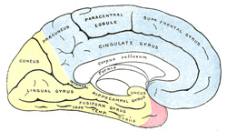
| 8. The anterior cerebral artery (a. cerebri anterior) (Figs. 516, 517, 518) arises from the internal carotid, at the medial extremity of the lateral cerebral fissure. It passes forward and medialward across the anterior perforated substance, above the optic nerve, to the commencement of the longitudinal fissure. Here it comes into close relationship with the opposite artery, to which it is connected by a short trunk, the anterior communicating artery. From this point the two vessels run side by side in the longitudinal fissure, curve around the genu of the corpus callosum, and turning backward continue along the upper surface of the corpus callosum to its posterior part, where they end by anastomosing with the posterior cerebral arteries. | 26 |
| | | Branches.—In its course the anterior cerebral artery gives off the following branches: | 27 |
| Antero-medial Ganglionic. |
|
Anterior. |
|
Posterior. |
|
| Inferior. |
|
Middle. |
|
|
| The Antero-medial Ganglionic Branches are a group of small arteries which arise at the commencement of the anterior cerebral artery; they pierce the anterior perforated substance and lamina terminalis, and supply the rostrum of the corpus callosum, the septum pellucidum, and the head of the caudate nucleus. The inferior branches, two or three in number, are distributed to the orbital surface of the frontal lobe, where they supply the olfactory lobe, gyrus rectus, and internal orbital gyrus. The anterior branches supply a part of the superior frontal gyrus, and send twigs over the edge of the hemisphere to the superior and middle frontal gyri and upper part of the anterior central gyrus. The middle branches supply the corpus callosum, the cingulate gyrus, the medial surface of the superior frontal gyrus, and the upper part of the anterior central gyrus. The posterior branches supply the precuneus and adjacent lateral surface of the hemisphere. | 28 |
 |
FIG. 516– The arteries of the base of the brain. The tempora pole of the cerebrum and a portion of the cerebellar hemisphere have been removed on the right side. (See enlarged image) | | |
| The Anterior Communicating Artery (a. communicans anterior) connects the two anterior cerebral arteries across the commencement of the longitudinal fissure. Sometimes this vessel is wanting, the two arteries joining together to form a single trunk, which afterward divides; or it may be wholly, or partially, divided into two. Its length averages about 4 mm., but varies greatly. It gives off some of the antero-medial ganglionic vessels, but these are principally derived from the anterior cerebral. | 29 |
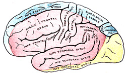
| 9. The middle cerebral artery (a. cerebri media) (Figs. 516, 517), the largest branch of the internal carotid, runs at first lateralward in the lateral cerebral or Sylvian fissure and then backward and upward on the surface of the insula, where it divides into a number of branches which are distributed to the lateral surface of the cerebral hemisphere. | 30 |
| | | Branches.—The branches of this vessel are the: | 31 |
| Antero-lateral Ganglionic. |
|
Ascending Parietal. |
| Inferior Lateral Frontal. |
|
Parietotemporal. |
| Ascending Frontal. |
|
Temporal. | |
 |
FIG. 517– Outer surface of cerebral hemisphere, showing areas supplied by cerebral arteries. (See enlarged image) | | |
 |
FIG. 518– Medial surface of cerebral hemisphere, showing areas supplied by cerebral arteries. (See enlarged image) | | |
| The Antero-lateral Ganglionic Branches, a group of small arteries which arise at the commencement of the middle cerebral artery, are arranged in two sets: one, the internal striate, passes upward through the inner segments of the lentiform nucleus, and supplies it, the caudate nucleus, and the internal capsule; the other, the external striate, ascends through the outer segment of the lentiform nucleus, and supplies the caudate nucleus and the thalamus. One artery of this group is of larger size than the rest, and is of special importance, as being the artery in the brain most frequently ruptured; it has been termed by Charcot the artery of cerebral hemorrhage. It ascends between the lentiform nucleus and the external capsule, and ends in the caudate nucleus. The inferior lateral frontal supplies the inferior frontal gyrus (Broca’s convolution) and the lateral part of the orbital surface of the frontal lobe. The ascending frontal supplies the anterior central gyrus. The ascending parietal is distributed to the posterior central gyrus and the lower part of the superior parietal lobule. The parietotemporal supplies the supramarginal and angular gyri, and the posterior parts of the superior and middle temporal gyri. The temporal branches, two or three in number, are distributed to the lateral surface of the temporal lobe. | 32 |
| 10. The posterior communicating artery (a. communicans posterior) (Fig. 516) runs backward from the internal carotid, and anastomoses with the posterior cerebral, a branch of the basilar. It varies in size, being sometimes small, and occasionally so large that the posterior cerebral may be considered as arising from the internal carotid rather than from the basilar. It is frequently larger on one side than on the other. From its posterior half are given off a number of small branches, the postero-medial ganglionic branches, which, with similar vessels from the posterior cerebral, pierce the posterior perforated substance and supply the medial surface of the thalami and the walls of the third ventricle. | 33 |
| 11. The anterior choroidal (a. chorioidea; choroid artery) is a small but constant branch, which arises from the internal carotid, near the posterior communicating artery. Passing backward and lateralward between the temporal lobe and the cerebral peduncle, it enters the inferior horn of the lateral ventricle through the choroidal fissure and ends in the choroid plexus. It is distributed to the hippocampus, fimbria, tela chorioidea of the third ventricle, and choroid plexus. | 34 |
|
|


