| Henry Gray (1821–1865). Anatomy of the Human Body. 1918. |
| |
| 13. Development of the Body Cavities |
| |
| In the human embryo described by Peters the mesoderm outside the embryonic disk is split into two layers enclosing an extra-embryonic cœlom; there is no trace of an intra-embryonic cœlom. At a later stage four cavities are formed within the embryo, viz., one on either side within the mesoderm of the pericardial area, and one in either lateral mass of the general mesoderm. All these are at first independent of each other and of the extra-embryonic celom, but later they become continuous. The two cavities in the general mesoderm unite on the ventral aspect of the gut and form the pleuro-peritoneal cavity, which becomes continuous with the remains of the extra-embryonic celom around the umbilicus; the two cavities in the pericardial area rapidly join to form a single pericardial cavity, and this from two lateral diverticula extend caudalward to open into the pleuro-peritoneal cavity (Fig. 54). | 1 |
 |
FIG. 54– Figure obtained by combining several successive sections of a human embryo of about the fourth week (From Kollmann.) The upper arrow is in the pleuroperitoneal opening, the lower in the pleuropericardial. (See enlarged image) | | |
| Between the two latter diverticula is a mass of mesoderm containing the ducts of Cuvier, and this is continuous ventrally with the mesoderm in which the umbilical veins are passing to the sinus venosus. A septum of mesoderm thus extends across the body of the embryo. It is attached in front to the body-wall between the pericardium and umbilicus; behind to the body-wall at the level of the second cervical segment; laterally it is deficient where the pericardial and pleuro-peritoneal cavities communicate, while it is perforated in the middle line by the foregut. This partition is termed the septum transversum, and is at first a bulky plate of tissue. As development proceeds the dorsal end of the septum is carried gradually caudalward, and when it reaches the fifth cervical segment muscular tissue with the phrenic nerve grows into it. It continues to recede, however, until it reaches the position of the adult diaphragm on the bodies of the upper lumbar vertebræ. The liver buds grow into the septum transversum and undergo development there. | 2 |
| The lung buds meantime have grown out from the fore-gut, and project laterally into the forepart of the pleuro-peritoneal cavity; the developing stomach and liver are imbedded in the septum transversum; caudal to this the intestines project into the back part of the pleuro-peritoneal cavity (Fig. 55). Owing to the descent of the dorsal end of the septum transversum the lung buds come to lie above the septum and thus pleural and peritoneal portions of the pleuro-peritoneal cavity (still, however, in free communication with one another) may be recognized; the pericardial cavity opens into the pleural part. | 3 |
 |
FIG. 55– Upper part of celom of human embryo of 6.8 mm., seen from behind. (From model by Piper.) (See enlarged image) | | |
| The ultimate separation of the permanent cavities from one another is effected by the growth of a ridge of tissue on either side from the mesoderm surrounding the duct of Cuvier (Figs. 54, 55). The front part of this ridge grows across and obliterates the pleuro-pericardial opening; the hinder part grows across the pleuro-peritoneal opening. | 4 |
 |
FIG. 56– Diagram of transverse section through rabbit embryo. (After Keith.) (See enlarged image) | | |
| With the continued growth of the lungs the pleural cavities are pushed forward in the body-wall toward the ventral median line, thus separating the pericardium from the lateral thoracic walls (Fig. 53). The further development of the peritoneal cavity has been described with the development of the digestive tube (page 168 et seq.). | 5 |
 |
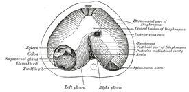
FIG. 57– The thoracic aspect of the diaphragm of a newly born child in which the communication between the peritoneum and pleura has not been closed on the left side; the position of the opening is marked on the right side by the spinocostal hiatus. (After Keith.) (See enlarged image) | | |
|
|


