| Henry Gray (1821–1865). Anatomy of the Human Body. 1918. |
| |
| 4a. The Pericardium |
| |
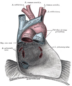
| The pericardium (Fig. 489) is a conical fibro-serous sac, in which the heart and the roots of the great vessels are contained. It is placed behind the sternum and the cartilages of the third, fourth, fifth, sixth, and seventh ribs of the left side, in the mediastinal cavity. | 1 |
| In front, it is separated from the anterior wall of the thorax, in the greater part of its extent, by the lungs and pleuræ; but a small area, somewhat variable in size, and usually corresponding with the left half of the lower portion of the body of the sternum and the medial ends of the cartilages of the fourth and fifth ribs of the left side, comes into direct relationship with the chest wall. The lower extremity of the thymus, in the child, is in contact with the front of the upper part of the pericardium. Behind, it rests upon the bronchi, the esophagus, the descending thoracic aorta, and the posterior part of the mediastinal surface of each lung. Laterally, it is covered by the pleuræ, and is in relation with the mediastinal surfaces of the lungs; the phrenic nerve, with its accompanying vessels, descends between the pericardium and pleura on either side. | 2 |
 |
FIG. 489– Posterior wall of the pericardial sac, showing the lines of reflection of the serous pericardium on the great vessels. (See enlarged image) | | |
| | | Structure of the Pericardium.—Although the pericardium is usually described as a single sac, an examination of its structure shows that it consists essentially of two sacs intimately connected with one another, but totally different in structure. The outer sac, known as the fibrous pericardium, consists of fibrous tissue. The inner sac, or serous pericardium, is a delicate membrane which lies within the fibrous sac and lines its walls; it is composed of a single layer of flattened cells resting on loose connective tissue. The heart invaginates the wall of the serous sac from above and behind, and practically obliterates its cavity, the space being merely a potential one. | 3 |
| The fibrous pericardium forms a flask-shaped bag, the neck of which is closed by its fusion with the external coats of the great vessels, while its base is attached to the central tendon and to the muscular fibers of the left side of the diaphragm. In some of the lower mammals the base is either completely separated from the diaphragm or joined to it by some loose areolar tissue; in man much of its diaphragmatic attachment consists of loose fibrous tissue which can be readily broken down, but over a small area the central tendon of the diaphragm and the pericardium are completely fused. Above, the fibrous pericardium not only blends with the external coats of the great vessels, but is continuous with the pretracheal layer of the deep cervical fascia. By means of these upper and lower connections it is securely anchored within the thoracic cavity. It is also attached to the posterior surface of the sternum by the superior and inferior sternopericardiac ligaments; the upper passing to the manubrium, and the lower to the xiphoid process. | 4 |
| The vessels receiving fibrous prolongations from this membrane are: the aorta, the superior vena cava, the right and left pulmonary arteries, and the four pulmonary veins. The inferior vena cava enters the pericardium through the central tendon of the diaphragm, and receives no covering from the fibrous layer. | 5 |
| The serous pericardium is, as already stated, a closed sac which lines the fibrous pericardium and is invaginated by the heart; it therefore consists of a visceral and a parietal portion. The visceral portion, or epicardium, covers the heart and the great vessels, and from the latter is continuous with the parietal layer which lines the fibrous pericardium. The portion which covers the vessels is arranged in the form of two tubes. The aorta and pulmonary artery are enclosed in one tube, the arterial mesocardium. The superior and inferior venæ cavæ and the four pulmonary veins are enclosed in a second tube, the venous mesocardium, the attachment of which to the parietal layer presents the shape of an inverted U. The cul-de-sac enclosed between the limbs of the U lies behind the left atrium and is known as the oblique sinus, while the passage between the venous and arterial mesocardia—i.e., between the aorta and pulmonary artery in front and the atria behind—is termed the transverse sinus. | 6 |
| | | The Ligament of the Left Vena Cava.—Between the left pulmonary artery and subjacent pulmonary vein is a triangular fold of the serous pericardium; it is known as the ligament of the left vena cava (vestigial fold of Marshall). It is formed by the duplicature of the serous layer over the remnant of the lower part of the left superior vena cava (duct of Cuvier), which becomes obliterated during fetal life, and remains as a fibrous band stretching from the highest left intercostal vein to the left atrium, where it is continuous with a small vein, the vein of the left atrium (oblique vein of Marshall), which opens into the coronary sinus. | 7 |
| The arteries of the pericardium are derived from the internal mammary and its musculophrenic branch, and from the descending thoracic aorta. | 8 |
| The nerves of the percardium are derived from the vagus and phrenic nerves, and the sympathetic trunks. | 9 |
|
|


